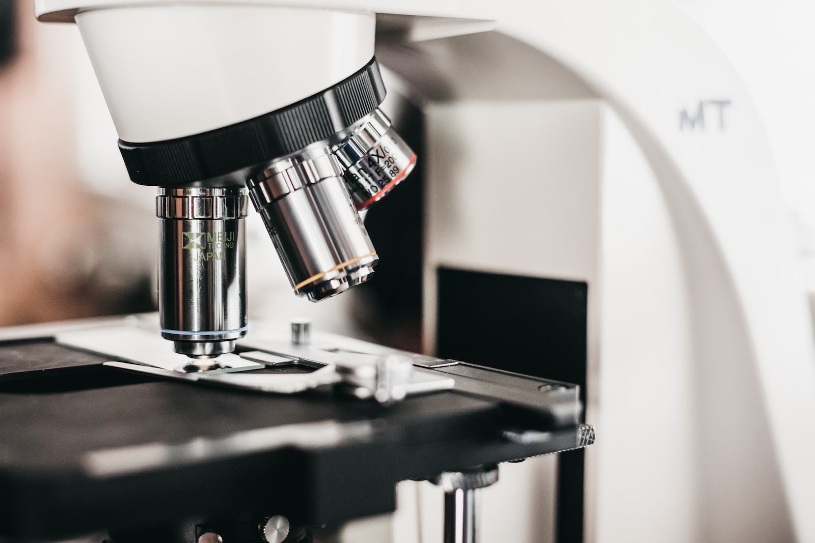
A semen microscope is a great way to examine sperm and see the quality of it. This sperm microscope has a 400x magnification, and LED light is cool enough to not heat up the sperm and kill them. Coarse and fine focusing make it easy to get a clear image.
Head
A sperm microscope is used by andrologists to analyze semen samples for fertility testing. The best sperm analysis microscopes feature a heated stage to keep the sample warm and phase-contrast technology for contrasting stains without harming the cells. Many also allow you to capture images or videos for documentation and training purposes.
A healthy sperm has an oval head and long tails. Abnormal sperm may have enlarged heads, narrow or crooked tails or double tails. This can be due to a low DNA fragmentation index (DFI), which is a marker of male infertility.
The head of a sperm is covered with a cap called the acrosomal cap, anterior nuclear cap or galea capitis. The acrosomal cap helps to protect the sperm from oxidation and provides the necessary energy for sperm motility. The neck of the sperm is comprised of a funnel-shaped structure called the basal body that unites the head with the middle piece. It is also where the acrosome is located, which is where the sperm’s spermicide chemicals are stored. The acrosomal cap also contains receptors for sperm to bind with ovarian lining cells.
Neck
You can see the neck of a sperm under a microscope, as well as different parts of the head and tail. You can use a labelled diagram to help you identify these structures. Normally, healthy sperm have a smooth head and neck, but you can also find sperm with defects such as crooked heads or double tails.
The neck of a sperm is a narrow structure that connects the head to the middle piece. It contains a centrally located centriole and a funnel-shaped basal body that helps union the head with the middle piece. The neck also has coarse outer fibres that join with the coarse middle part of the sperm.
Sperm are important for reproduction, and evaluating the number and quality of sperm is essential to livestock breeding programs. A specialised microscope is needed to examine sperm samples, and it’s important to choose one with the right magnification for this application. A semen analysis microscope with phase contrast microscopy is ideal, as it turns the tiny phases shifts in light passing through a sample into visible brightness variations. This improves contrast and is safe for the specimens.
Middle Piece
The middle region of sperm contains a compact helical mass of mitochondria. These mitochondria provide the sperm with energy for movement and fertilization of the egg. This middle piece also contains nine peripheral doublets microtubules. These microtubules form a complex axial filament that extends from the middle to the tail.
The tail of sperm is long and whip-like. It is a vital part of sperm motility and facilitates the penetration of the egg’s outer surface. The tail also has a plasma membrane that protects the sperm from damage.
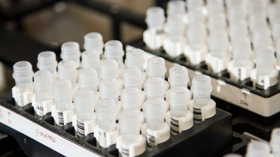
A sperm microscope, or semen microscope, is used to identify and count spermatozoa. These microscopic tools use depression slides and cover slips to examine sperm samples. They are available in a variety of magnifications, including 400x. A good sperm microscope should also have a heated stage or be capable of adding one. This is necessary to ensure that the sperm samples stay alive and do not perish during inspection.
Normal sperm are characterized by their head size, head DNA content, midpiece appearance and tail structure. Abnormal sperm, however, can decrease fertility by impairing their ability to penetrate and fertilize the woman’s egg. Therefore, it is important to have a proper semen analysis performed by an experienced professional.
Principal Piece
During motility evaluation, the sperm’s ability to move and swim is important. A normal light microscope won’t be able to see sperm moving properly, so you need one that is specifically designed for this purpose. This specialized sperm microscope has LED lights that can provide up to 400x magnification, and the lenses have coarse and fine focusing, allowing you to get a clear image. It also has a stage that can be heated to keep your samples warm during observation, as without warmth they will quickly perish.
The sperm head, neck, and middle piece all have a dark color when viewed under the microscope. The acrosomal cap and the middle part of the tail have a lighter color. The acrosomal cap has a thick structure that helps to protect the sperm from harmful substances in the environment. You can identify all the different parts of the sperm under the microscope using the routine spermac stain, which gives each of them a unique colour. You can also identify the other structures in a seminiferous tubule or epididymis slide, like Leydig cells and interstitial connective tissue.
Tail
Sperm morphology, a term that refers to the shape of the sperm, can impact its ability to penetrate the egg and fertilize it. This shape is assessed during a routine semen analysis, which includes tests for motility and concentration.
The tail piece of a sperm is composed of an axial filament that is connected to nine columns of dense outer fibres. These outer fibres wrap around the axoneme and form a mitochondrial sheath. The axial filament also fuses with the middle piece’s structural protein and forms an asymmetrical endpiece.
The asymmetrical end of the middle piece is called the acrosome cap, and it covers between 40-70% of the head. The acrosome is surrounded by a sheath that protects the head and neck from the ionic and chromosomal environment in which sperm swim. These structures are visible under a light microscope when the sperm is stained with a routine dye called spermac stain, which colors different parts of the sperm based on their metabolic activity. With this technique, the head, neck, acrosome, and middle and principal pieces can all be seen clearly.